Addiction and Mitochondria
by Kenneth Anderson, M.A.
A recent paper by Jun Gao and colleagues found that methamphetamine, morphine, and heroin result in high energy demands of the neurons which release dopamine into the nucleus accumbens. These high energy demands force the mitochondria of these cells to kick into overdrive to produce enough energy to keep up with these demands. The researchers found that blocking these mitochondria from kicking into overdrive could block the addictive properties of these drugs without blocking natural rewards such as the reward from food. This is a potentially important discovery, since blocking dopamine itself blocks natural rewards and can lead to anhedonia (loss of pleasure) and motivational blunting. However, the researchers also found that cocaine does not kick the mitochondria into overdrive but instead works by a very different mechanism than the other drugs mentioned above. In this blog post, I will go over the findings of this paper. However, let’s first review how neurotransmission works.
A multipolar neuron consists of a cell body, several dendrites, and an axon. The dendrites of a multipolar neuron act as electrical inputs, and the axon functions as an electrical output. Multipolar neurons are constantly receiving electrical signals through their dendrites. When the sum of these electrical inputs from the dendrites reaches a certain critical level, the neuron fires; i.e., the neuron sends an electrical signal down its axon. The resting potential of the neuron is about -70 millivolts. The threshold potential, which causes the neuron to fire, is around -55 millivolts. Multipolar neurons are the most common type of neuron found in humans. Although a few other types of neurons exist, these do not concern us here.
When the neuron fires, it sends an electrical signal down the axon. The axon ends in a number of axon terminals, each of which can form half of a synapse. A synapse consists of a bulb at the end of the axon terminal (known as a bouton), the dendrite of another neuron, and the gap between them. When the electrical signal reaches the bulb at the end of the axon, it causes the bulb to release certain chemicals known as neurotransmitters. These neurotransmitters cross the synaptic gap and bind to receptors on the dendrite of the other neuron, creating an electrical signal which travels toward the cell body of the neuron to be summed up with the electrical signals from this neuron’s other dendrites. The electrical signals created when the neurotransmitter binds to the receptor can be either more electrically positive or more electrically negative, depending on what the neurotransmitter is. Positive signals are called excitatory, since they increase the likelihood of the neuron firing. Negative signals are called inhibitory since they decrease the likelihood of the neuron firing. All of this happens very rapidly. The synaptic gap is typically about 20 nanometers wide, and the time it takes for the neurotransmitters to cross the synaptic gap is typically a fraction of a millisecond.
The neurons we are interested in are neurons with their cell bodies located in the ventral tegmental area (VTA) of the brain, which is located in the lower (ventral) part of the midbrain. These neurons send out axons to the nucleus accumbens, the prefrontal cortex, and a number of other parts of the brain. We are particularly interested in the axons which go from the ventral tegmental area (VTA) to the nucleus accumbens, because these neurons are an important part of the reward pathway, which is involved in habit formation, including the formation of addictions. The nucleus accumbens is located in the forebrain.
The neurons which send out axons from the ventral tegmental area (VTA) to the nucleus accumbens are dopamine-releasing neurons. Like many other neurons in the brain, these neurons are constantly firing at a relatively slow baseline rate; they fire about two to five times per second (i.e., 2 to 5 Hertz). Think of this as similar to the idling of a car engine. Certain stimuli, such as a food reward, can cause the neuron to change from the slower tonic firing rate to the faster phasic firing rate: 15 to 30 times per second (Hertz) for rodent dopamine neurons. Think of this as like putting your car in gear and stepping on the gas. The faster firing rate means that more of the neurotransmitter, in this case dopamine, is released into the synaptic gap.
Now let’s take a closer look at what is happening in that bulb (bouton) at the end of the axon terminal, which is what releases the neurotransmitter molecules into the synaptic gap. Inside this bulb are many little sacks, known as vesicles, which are filled with neurotransmitter molecules. When the electrical signal travels down the axon and reaches the bulb, some of these little sacks move towards the synaptic gap, fuse with the cell membrane of the bulb, burst open, and release their neurotransmitter molecules into the synaptic gap. Some of the neurotransmitter molecules bind to the receptors on the dendrite of the neuron which is receiving the message; however, most of the neurotransmitter molecules wind up floating in the synaptic gap. That’s when neurotransmitter reuptake transporters go to work. Neurotransmitter reuptake transporters are tiny cylinders made of protein and embedded in the cell membrane of the axon bulb. These neurotransmitter transporters vacuum up the stray neurotransmitter molecules floating in the synaptic gap and return them to the inside of the axon bulb, where they are repackaged into sacks, ready to repeat the cycle all over again. This process of returning the neurotransmitter molecules into the axon bulb is known as “reuptake.” Although the neuron is constantly synthesizing new neurotransmitter molecules to replace the old ones, reuptake is responsible for replacing most of the neurotransmitters which have been released. This process is illustrated in Figure 1.

When a reward such as food is present, many of the neurons going from the ventral tegmental area (VTA) to the nucleus accumbens switch from slow, tonic firing to more rapid, phasic firing, and the result is that more dopamine is released from these neurons into the nucleus accumbens. However, the dopamine increase caused by food reward is not huge; one study using rats found a dopamine increase of 37% in the nucleus accumbens as a result of food reward. On the other hand, cocaine, amphetamine and methamphetamine all cause huge increases in dopamine released into the nucleus accumbens–the previously mentioned study showed that amphetamine and cocaine both led to an increase of five times (500%) baseline, and some studies have found similar or greater dopamine increases caused by methamphetamine.
Additionally, methamphetamine causes the dopamine reuptake transporter to function in reverse. Instead of sucking up dopamine molecules and returning them into the axon bulb, the dopamine reuptake transporter is now spraying the dopamine molecules out of the axon bulb and into the synaptic gap. The high-intensity dopamine activity and sustained dopamine release place a much greater energy demand on the neuron than usual. (Methamphetamine also affects the vesicular monoamine transporter 2 [VMAT2], but I won’t go into that in this post.)
Cocaine also greatly increases the amount of dopamine in the synaptic gap, but the mechanism is very different from that of methamphetamine. Cocaine acts as a dopamine reuptake inhibitor. Most of us are familiar with selective serotonin reuptake inhibitors (SSRIs) such as Prozac. SSRIs work by blocking the reuptake of serotonin by blocking the serotonin reuptake transporter, leaving a greater concentration of serotonin in the synaptic gap. Cocaine does the same thing with dopamine. In fact, cocaine not only blocks reuptake of dopamine, but also of norepinephrine and serotonin, making it a nonselective reuptake inhibitor. However, it is the effect of cocaine on dopamine that makes the cocaine high similar to the methamphetamine high. But cocaine does not create the same energy demand in the neuron that methamphetamine does.
Like methamphetamine, heroin and morphine also create a large increase in the amount of dopamine which the neurons going from the ventral tegmental area (VTA) to the nucleus accumbens release into the synaptic gap; however, the mechanism is an indirect one. There are GABA-releasing neurons which connect to the dopamine-releasing neurons going from the ventral tegmental area (VTA) to the nucleus accumbens. GABA is an inhibitory neurotransmitter, which means it acts like a brake on these dopamine-releasing neurons. When opioids such as morphine bind to the receptors on these GABA-releasing neurons, they greatly reduce the amount of GABA released. This is similar to taking your foot off the brake. The dopamine-releasing neurons now have their brakes turned off. I have discussed this mechanism in an earlier blog post.
This brings us back to the paper by Jun Gao and colleagues that we mentioned at the beginning of this article. As I said above, these researchers found that the energy demands of dopamine-releasing neurons when under the influence of methamphetamine or morphine kick the mitochondria into overdrive.
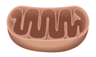
The mitochondrion is nicknamed “the powerhouse of the cell.” The mitochondria power the cell by synthesizing ATP and releasing it into the cytoplasm. The mitochondrion is comprised of an outer membrane, an inner membrane, and a core known as the matrix. Normally, the mitochondrion produces ATP through a process involving the Krebs cycle. Calcium ions cannot freely cross the inner membrane to enter the matrix. A special transporter known as the mitochondrial calcium uniporter (MCU) is required to transport calcium ions across the inner membrane and into the matrix. In times of high energy demand, the MCU begins transporting a lot of calcium ions across the inner membrane to greatly increase the production of ATP. And as we saw above, morphine and methamphetamine create high energy demands on the dopamine-releasing neurons we were discussing.
Jun Gao and colleagues found that if they turned the MCU off, mice no longer exhibited addiction-like behaviors in relation to morphine or methamphetamine. However, this was not the case with cocaine.
One of the standard animal models of addiction in humans is conditioned place preference (CPP) in animals. What this means is that if the addictive drug is administered to an animal in a certain location, the animal starts to prefer hanging out in that location even when not receiving the drug. When the researchers turned off the MCU prior to giving the mice methamphetamine, they found that the mice developed no preference for the place where they received methamphetamine. The same was true with heroin and morphine. However, turning off the MCU had no effect in blocking conditioned place preference when the mice received cocaine. The mice still developed a preference for the location where they received cocaine. This suggests that the MCU is not involved in cocaine addiction, whereas the MCU is essential for developing an addiction to methamphetamine or opiates.
The researchers also investigated whether addiction-like behaviors could be reinstated in mice. The mice were first habituated to methamphetamine, morphine, or cocaine. The behavior was then extinguished. Next, the MCU was turned off. In the mice that received methamphetamine or morphine, a new dose of the drug failed to reinstate addiction-like behaviors. Cues associated with the drug, such a light which had been flashed when the mice were given the drug, also failed to reinstate addiction-like behaviors. However, this was not true of cocaine. Mice which had been habituated to cocaine immediately reinstated their addiction-like behaviors, despite the fact that the MCU had been turned off.
This suggests that drugs like berberine, which can turn off the MCU, could be useful in treating humans with addictions to opiates or methamphetamine, but not cocaine. Clinical trials of berberine would be needed to show that it was safe and effective.